A) supinated.
B) pronated.
C) lateral.
D) in 30-degree medial rotation.
Correct Answer

verified
Correct Answer
verified
Multiple Choice
For a lateral projection of the hand,the central ray is directed to enter the:
A) second digit MCP joint.
B) PIP joint.
C) distal PIP joint.
D) midmetacarpal area.
Correct Answer

verified
Correct Answer
verified
Multiple Choice
The bone part shown in this figure is the: 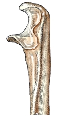
A) distal ulna.
B) proximal ulna.
C) distal radius.
D) proximal radius.
Correct Answer

verified
Correct Answer
verified
Multiple Choice
For a PA projection of the second digit,the central ray is directed to the:
A) distal interphalangeal joint.
B) proximal interphalangeal joint.
C) metacarpophalangeal joint.
D) carpometacarpal joint.
Correct Answer

verified
Correct Answer
verified
Multiple Choice
How many phalanges are there in the thumb?
A) One
B) Two
C) Three
D) Four
Correct Answer

verified
Correct Answer
verified
Multiple Choice
What anatomic structure is shown in profile on an AP projection of the humerus?
A) Capitulum
B) Glenoid cavity
C) Greater tubercle
D) Lesser tubercle
Correct Answer

verified
Correct Answer
verified
Multiple Choice
Which projection of the first digit is demonstrated in the figure above? 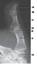
A) PA
B) PA oblique
C) Mediolateral
D) Lateromedial
Correct Answer

verified
Correct Answer
verified
Multiple Choice
What anatomy of the third digit is labeled as letter D in the figure above? 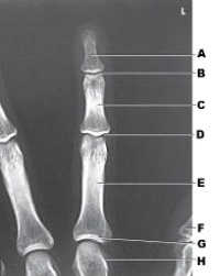
A) Distal IP joint
B) Proximal IP joint
C) Metacarpophalangeal joint
D) Carpometacarpal joint
Correct Answer

verified
Correct Answer
verified
Multiple Choice
If the IR and wrist are elevated for the PA axial projection of the wrist (Stecher method) ,the central ray orientation is:
A) perpendicular to the IR.
B) 20 degrees toward the elbow.
C) 20 degrees toward the hand.
D) variable according to the degree of IR/part elevation.
Correct Answer

verified
Correct Answer
verified
Multiple Choice
How many degrees is the elbow flexed for the lateral projection of the elbow?
A) 0
B) 45
C) 75
D) 90
Correct Answer

verified
Correct Answer
verified
Multiple Choice
What anatomy is labeled as letter A in the image below? 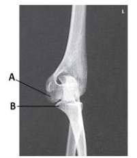
A) Capitulum
B) Lateral epicondyle of humerus
C) Trochlea
D) Coronoid process of ulna
Correct Answer

verified
Correct Answer
verified
Multiple Choice
What anatomy is labeled as letter E in the image below? 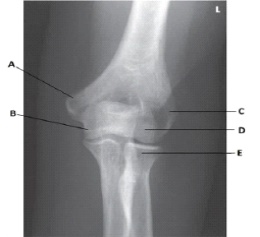
A) Radial head
B) Capitulum
C) Coronoid process of ulna
D) Trochlea
Correct Answer

verified
Correct Answer
verified
Multiple Choice
The bone part identified by the arrow in this figure is the: 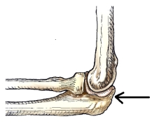
A) trochlea.
B) capitulum.
C) radial head.
D) olecranon process.
Correct Answer

verified
Correct Answer
verified
Multiple Choice
Which of the following is the largest carpal bone?
A) Capitate
B) Hamate
C) Scaphoid
D) Triquetrum
Correct Answer

verified
Correct Answer
verified
Multiple Choice
What anatomy is indicated by the arrow in this figure? 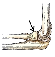
A) Trochlea
B) Capitulum
C) Radial head
D) Olecranon process
Correct Answer

verified
Correct Answer
verified
Multiple Choice
Which two of the following methods can be used to demonstrate the first CMC joint? (Select all that apply.)
A) Robert
B) Burman
C) Stecher
D) Norgaard
Correct Answer

verified
Correct Answer
verified
Showing 121 - 136 of 136
Related Exams