A) dura mater
B) epidural space
C) subarachnoid space
D) arachnoid mater
E) pia mater
Correct Answer

verified
Correct Answer
verified
Multiple Choice
The median nerve ________.
A) arises from both the medial and posterior cords of the brachial plexus
B) arises from both the medial and lateral cords of the brachial plexus
C) arises from the posterior cord of the brachial plexus
D) innervates the pronators of the forearm
E) is a major nerve of the cervical plexus
Correct Answer

verified
Correct Answer
verified
Multiple Choice
Spinal reflexes ________.
A) include monosynaptic reflexes only
B) involve only a single segment of the spinal cord
C) always transmit information to the brain for processing
D) include both monosynaptic and polysynaptic reflexes
E) do not include stretch reflexes
Correct Answer

verified
Correct Answer
verified
Multiple Choice
Figure 14.2
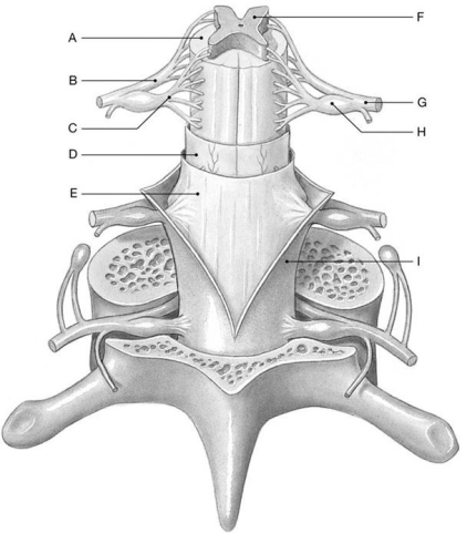 Using the above-referenced diagram of a posterior view of the spinal cord showing the meningeal layers, superficial landmarks, and distribution of gray and white matter, identify the specified labeled structure(s) in each of the following questions.
-Identify the structure(s) indicated by Label F.
A) Dorsal root
B) Ventral root
C) Dura mater
D) Gray matter
E) White matter
Correct Answer

verified
Correct Answer
verified
Multiple Choice
If you bump the "funny bone" which nerve function is interrupted?
A) median
B) radial
C) ulnar
D) axillary
E) musculocutaneous
Correct Answer

verified
Correct Answer
verified
Multiple Choice
The ________ arises from the posterior cord of the brachial plexus.
A) ulnar nerve
B) median nerve
C) radial nerve
D) musculocutaneous nerve
E) dorsal scapular nerve
Correct Answer

verified
Correct Answer
verified
Multiple Choice
Figure 14.7
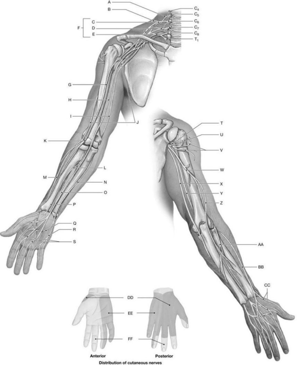 Using the above-referenced diagrams of the anterior and posterior views of the upper limb, identify the specified labeled structure(s) in each of the following questions.
-Identify the structure(s) indicated by Label EE.
A) Radial nerve
B) Musculocutaneous nerve
C) Ulnar nerve
D) Axillary nerve
E) Median nerve
Correct Answer

verified
Correct Answer
verified
Multiple Choice
All of the following are true of fiber tracts in the spinal cord except ________.
A) all axons within a tract relay information in the same direction
B) each tract carries sensory or motor information, but not both
C) axons of a single tract are relatively uniform in diameter and conduction speed
D) the tracts are randomly located with respect to the type of information carried
E) axons of a single tract are relatively uniform with respect to myelination
Correct Answer

verified
Correct Answer
verified
Multiple Choice
Figure 14.8
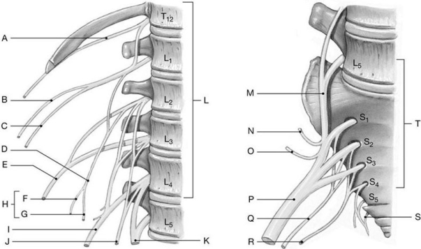 Using the above-referenced diagrams of the lumbar and sacral plexuses, identify the specified labeled structure(s) in each of the following questions.
-Identify the structure(s) indicated by Label D.
A) Iliohypogastric nerve
B) Genitofemoral nerve
C) Sciatic nerve
D) Pudendal nerve
E) Ilio-inguinal nerve
Correct Answer

verified
Correct Answer
verified
Multiple Choice
Figure 14.3
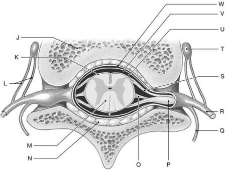 Using the above-referenced diagram of a sectional view through the spinal cord and meninges showing the peripheral distribution of the spinal nerves, identify the specified labeled structure(s) in each of the following questions.
-Identify the structure(s) indicated by Label P.
A) Rami communicantes
B) Dorsal root ganglion
C) Ventral ramus
D) Dorsal ramus
E) Ventral root
Correct Answer

verified
Correct Answer
verified
Multiple Choice
The short head of the biceps femoris and the tibialis anterior muscles are innervated by the ________ nerve.
A) iliohypogastric
B) pudendal
C) fibular
D) inferior gluteal
E) lateral femoral cutaneous
Correct Answer

verified
Correct Answer
verified
True/False
The brachial plexus is composed of cutaneous and muscular branches of the ventral rami of spinal nerves C1-C4.
Correct Answer

verified
Correct Answer
verified
Multiple Choice
The ________ nerve of the lumbar plexus is formed by ventral rami of T12 and L1, and innervates the internal and external oblique muscles.
A) genitofemoral
B) lateral femoral cutaneous
C) ilioinguinal
D) subcostal
E) iliohypogastric
Correct Answer

verified
Correct Answer
verified
Multiple Choice
Figure 14.1
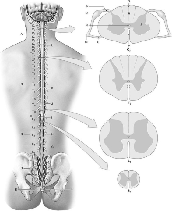 Using the above- referenced diagrams of the superficial anatomy and orientation of the adult spinal cord (posterior view) and inferior views of cross-sections through representative segments of the spinal cord, showing the arrangement of gray and white matter, identify the specified labeled structure(s) in each of the following questions.
-Identify the structure(s) indicated by Label O.
A) Ventral root
B) Dorsal root ganglion
C) Central canal
D) White matter
E) Anterior median fissure
Correct Answer

verified
Correct Answer
verified
Multiple Choice
Figure 14.2
 Using the above-referenced diagram of a posterior view of the spinal cord showing the meningeal layers, superficial landmarks, and distribution of gray and white matter, identify the specified labeled structure(s) in each of the following questions.
-Identify the structure(s) indicated by Label H.
A) White matter
B) Gray matter
C) Arachnoid mater
D) Dorsal root ganglion
E) Denticulate ligament
Correct Answer

verified
Correct Answer
verified
Multiple Choice
The largest nerve in the body that is formed from L4-S3 is the ________.
A) tibial nerve
B) pudendal nerve
C) sciatic nerve
D) fibular nerve
E) superior gluteal nerve
Correct Answer

verified
Correct Answer
verified
Multiple Choice
Figure 14.4
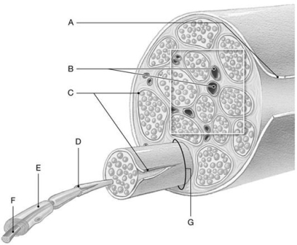 Using the above-referenced diagram of a typical peripheral nerve and its connective tissue wrappings, identify the specified labeled structure(s) in each of the following questions.
-Identify the structure(s) indicated by Label B.
A) Schwann cell
B) Epineurium
C) Fascicle
D) Perineurium
E) Blood vessels
Correct Answer

verified
Correct Answer
verified
Multiple Choice
Figure 14.8
 Using the above-referenced diagrams of the lumbar and sacral plexuses, identify the specified labeled structure(s) in each of the following questions.
-Identify the structure(s) indicated by Label B.
A) Genitofemoral nerve
B) Ilio-inguinal nerve
C) Sciatic nerve
D) Iliohypogastric nerve
E) Pudendal nerve
Correct Answer

verified
Correct Answer
verified
Multiple Choice
Activation of a sensory neuron results in the conduction of action potentials into the spinal cord along a(n) ________.
A) efferent fiber
B) ventral ramus
C) ventral horn
D) afferent fiber
E) peripheral effector
Correct Answer

verified
Correct Answer
verified
Multiple Choice
Figure 14.4
 Using the above-referenced diagram of a typical peripheral nerve and its connective tissue wrappings, identify the specified labeled structure(s) in each of the following questions.
-Identify the structure(s) indicated by Label C.
A) Perineurium
B) Schwann cell
C) Epineurium
D) Blood vessels
E) Endoneurium
Correct Answer

verified
Correct Answer
verified
Showing 61 - 80 of 131
Related Exams